Best Disease: The Five Stages on Fundus, FAF, OCT and EOG
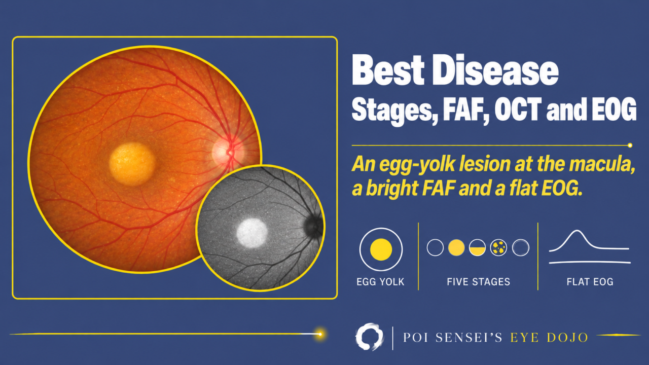
A child or young adult has a round, yellow lesion at the macula that looks like an egg yolk. Vision is often better than the fundus suggests. That is the typical picture of Best disease. This article shows how the lesion changes through its five stages, how it looks on fundus autofluorescence (FAF) and optical coherence tomography (OCT), and why the electro-oculogram (EOG) is the key test.
- A young patient has a round yellow lesion at the macula, often in both eyes.
- The lesion is intensely bright on FAF.
- OCT shows material under the retina.
- The EOG is abnormal, but the electroretinogram (ERG) is normal.
What is Best disease?
Best disease is an inherited macular dystrophy. Its full name is Best vitelliform macular dystrophy. It is the second most common macular dystrophy after Stargardt disease.
Inheritance is autosomal dominant. The gene is BEST1. It makes bestrophin, an ion channel in the retinal pigment epithelium (RPE) that is involved in chloride transport. When bestrophin does not work, RPE function fails, and yellow material builds up under the macula.
Severity varies widely, even within the same family[1].
Symptoms
Typical cases present before about age 20 with reduced vision or a central scotoma.
While the yellow "yolk" is intact, vision is often still good. Vision drops later, when the lesion breaks up and the macula becomes atrophic or scarred[1].
The five stages
Best disease is staged by its fundus appearance. The classic system has five stages[2].
Stage 1: Previtelliform
The fundus is normal, but the EOG is already abnormal. People with a BEST1 variant and a normal fundus also have an abnormal EOG.
Stage 2: Vitelliform
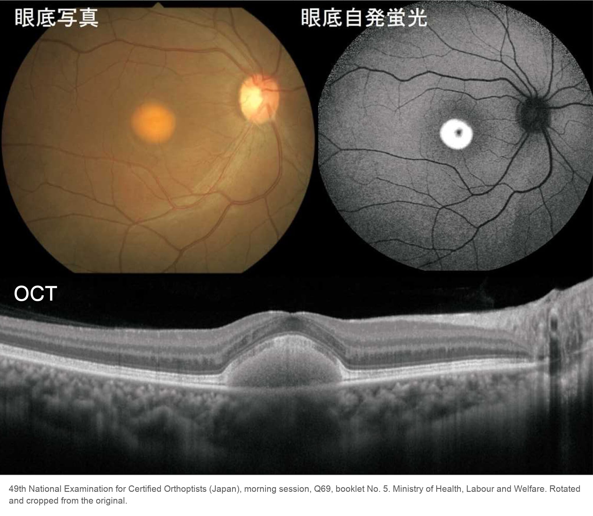
A round, sharply outlined yellow lesion appears at the macula, like the yolk of a sunny-side-up egg. It is 0.5 to 2 disc diameters across. The two eyes can be at different stages, and sometimes only one eye is affected at first. Vision is usually not affected yet[1].
The yellow material is thought to be lipofuscin. On OCT, it sits between the retina and the RPE as a dome. Lipofuscin is autofluorescent, so on FAF the lesion is intensely bright.
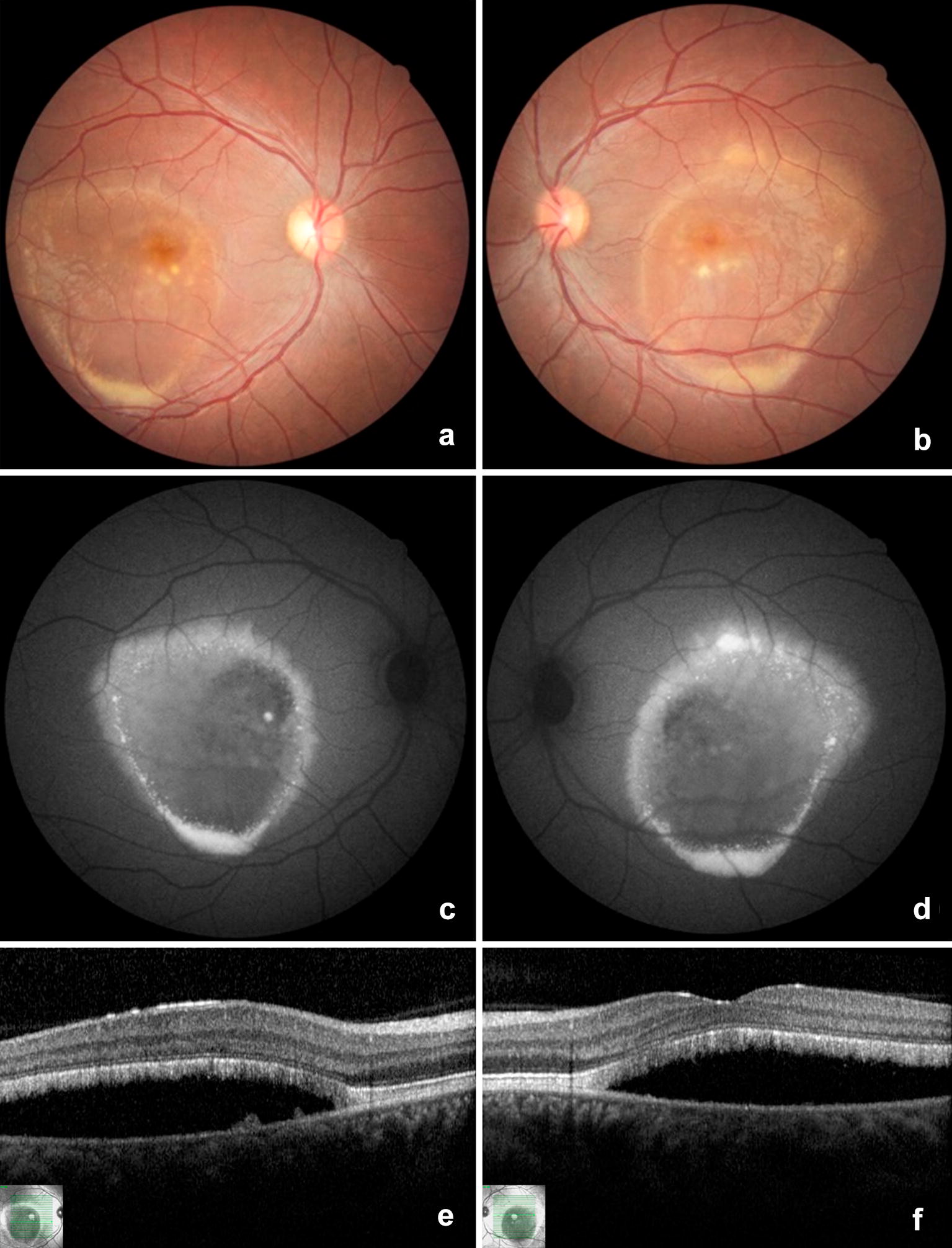
Stage 3: Pseudohypopyon
Part of the material breaks down, often around puberty[1]. The remaining yellow material settles to the bottom of the lesion, with a flat upper edge, like a hypopyon in the anterior chamber. OCT shows subretinal fluid above the settled material[3].
Stage 4: Vitelliruptive
The material breaks up into patches, like scrambled eggs, and vision drops[1]. As the deposit disappears, OCT shows the subretinal fluid more clearly.
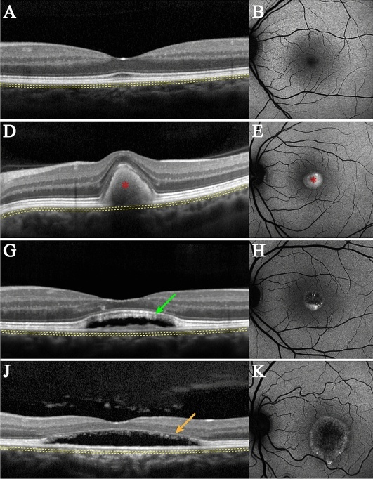
Stage 5: Atrophic
The yellow material is gone, leaving an atrophic area of RPE. FAF is now dark (hypoautofluorescent) in this area, and vision is clearly reduced. Some eyes end with a fibrotic scar instead[2].
EOG findings
The EOG is the key test for Best disease. It mainly reflects RPE function. It is usually abnormal at every stage, even before the fundus changes. The Arden ratio (the light peak divided by the dark trough, now called the LP:DT ratio) is typically below 1.5[1].
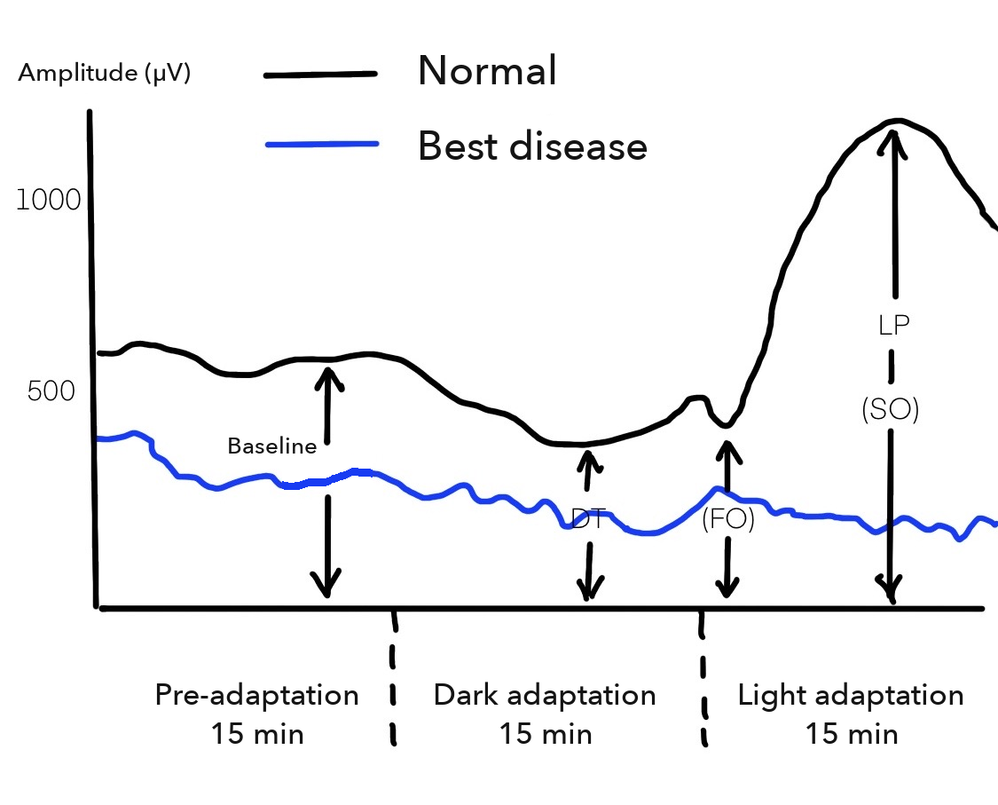
The full-field ERG is usually normal. An abnormal EOG with a normal ERG points to a problem in the RPE. The EOG article explains how the ratio is measured.
A few genetically confirmed eyes have a ratio above 1.5, so a normal EOG does not completely rule out Best disease. Genetic testing for BEST1 confirms the diagnosis[2].
Treatment
As with other macular dystrophies, there is no treatment for the disease itself.
Choroidal neovascularization (CNV) can develop and cause bleeding under the macula. It is treated with anti-VEGF injections. In a large UK series, about 1 in 6 patients had CNV during a mean follow-up of 8 years, and treated eyes kept better vision than untreated eyes[2].
Summary
- Best disease is an autosomal dominant macular dystrophy caused by BEST1.
- Five stages: normal fundus with an abnormal EOG → egg yolk → yolk settles with a level → scrambled egg → atrophy.
- The yellow material is bright on FAF and sits under the retina on OCT. Atrophy is dark on FAF.
- The EOG is usually abnormal at every stage, while the ERG is normal. Genetic testing confirms the diagnosis.
- There is no treatment for the dystrophy, but CNV is treated with anti-VEGF injections.
Figure credits
Vitelliform stage: reproduced from the 49th National Examination for Certified Orthoptists (Japan), morning session, question 69, published by the Ministry of Health, Labour and Welfare. Rotated and cropped from the original.
Pseudohypopyon stage: Figure 8 from de Souza CPR, Mello LGM, Gomez F, Morizot E. Int J Retina Vitreous 2019;5:6 (https://doi.org/10.1186/s40942-019-0156-0), licensed under CC BY 4.0. Unchanged.
Stages on OCT and autofluorescence: Figure 1 from Jauregui R, Parmann R, Nuzbrokh Y, Tsang SH, Sparrow JR. Sci Rep 2021;11:14300 (https://doi.org/10.1038/s41598-021-93316-3), licensed under CC BY 4.0. Cropped to remove the OCT angiography column (C, F, I, L).
EOG curves: drawn by the author.
References
[1] Salmon JF. Kanski’s Clinical Ophthalmology: A Systematic Approach. 10th ed. Elsevier; 2025. Chapter 15, Inherited retinal degenerations: Best vitelliform macular dystrophy.
[2] Laich Y, Georgiou M, Fujinami K, et al. Best vitelliform macular dystrophy natural history study report 1: clinical features and genetic findings. Ophthalmology. 2024;131(7):845-854. PMID 38278445.
[3] Jauregui R, Parmann R, Nuzbrokh Y, Tsang SH, Sparrow JR. Stage-dependent choriocapillaris impairment in Best vitelliform macular dystrophy characterized by optical coherence tomography angiography. Sci Rep. 2021;11(1):14300. PMID 34253754.
Which retinal layer makes which wave, the three abnormal patterns, and the dark-adaptation trap to check before you report “negative” — on one page, by email. One email when I publish something new, about once a month. Unsubscribe anytime.