Episcleritis or Scleritis? Pain, Phenylephrine and the Systemic Work-up
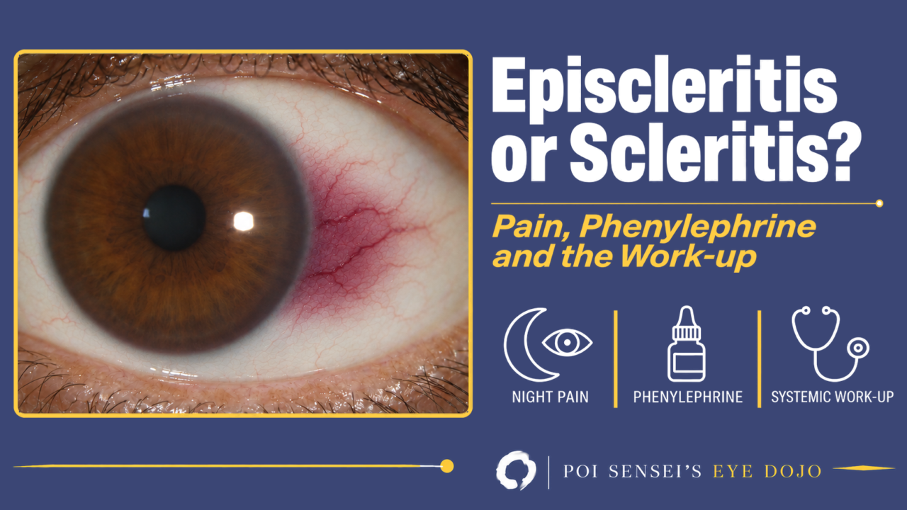
Episcleritis and scleritis both give a red eye with a patch of dilated vessels, and at first sight they can look alike, but the pain is different. Episcleritis is benign and settles on its own. Scleritis can destroy the eye, and it may be the first sign of a disease elsewhere in the body.
Pain is the first clue
Episcleritis causes mild discomfort or grittiness, and in up to half of patients no discomfort at all. The vision is normal.
Scleritis is usually painful. The pain is deep and boring, and it typically wakes the patient in the early hours of the morning. It can spread to the temple, brow or jaw and responds poorly to ordinary painkillers. The globe is tender when you press on it.
Few eye diseases cause pain like this, so a patient who says the eye pain woke them up should make you think of scleritis. The exception is scleromalacia perforans, a painless thinning of the sclera seen in long-standing rheumatoid arthritis.
How deep is the redness?
There are three layers of vessels on the front of the eye:
- Conjunctival vessels: the most superficial. They move with the conjunctiva.
- Superficial episcleral vessels: straight and radial. These are the most congested in episcleritis.
- Deep vascular plexus: in the outer layer of the sclera itself. This is where the congestion is greatest in scleritis.
Conjunctival injection is strongest in the periphery and fades toward the limbus. Ciliary injection is strongest at the limbus and comes from deeper vessels, so it points to deeper disease: acute angle closure, uveitis or scleritis.
At the slit lamp, four simple checks tell you the depth:
- Phenylephrine: 2.5% constricts the conjunctival vessels, and 10% also constricts the superficial episcleral vessels. The deep plexus does not blanch. Redness that stays after a drop of 10% phenylephrine is deep.
- Color: episcleritis is bright or salmon red. Scleritis has a purplish (violaceous) hue, best seen in daylight.
- A cotton bud: inflamed superficial episcleral vessels can be moved over the sclera. The vessels of the deep plexus cannot.
- The slit beam: an episcleral nodule is raised, but the sclera under it is flat. In nodular scleritis the sclera itself is raised.
Episcleritis
Episcleritis is common, benign and often recurrent. Most patients are young to middle-aged adults, and women may be affected more often.
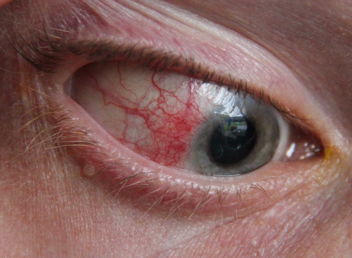
In this eye, the redness is limited to one sector beside the cornea, and the vessels are bright red.
There are two types:
- Simple episcleritis (about 75%): sectoral or diffuse redness, often in the interpalpebral area. It peaks within a day and fades over days to 3 weeks.
- Nodular episcleritis: a tender red nodule, almost always in the interpalpebral area. It lasts longer than the simple type.
An associated condition is found in about a third of patients. Ocular associations include rosacea, dry eye and contact lens wear, and systemic ones include rheumatoid arthritis. A single mild attack does not need an extensive work-up. Recurrent episcleritis should be investigated in the same way as scleritis.
Mild episcleritis needs no treatment. Cool compresses or refrigerated artificial tears help. If treatment is needed, a weak topical steroid four times a day for 1 to 2 weeks is usually enough; inflammation can rebound when the steroid is stopped. A topical NSAID is an alternative, though it may be less effective, and an oral NSAID is occasionally needed.
Scleritis is a systemic disease
Textbooks describe scleritis as a systemic disease that shows itself in the eye. Most cases are non-infectious, and an associated systemic disease is found in a third to a half of patients. Scleritis may be the first sign of that disease, so when you see scleritis, start by looking at the whole patient.
- Rheumatoid arthritis is the most common association. Scleritis in rheumatoid arthritis tends to run a more aggressive course.
- Granulomatosis with polyangiitis (formerly Wegener granulomatosis) often gives rapidly progressive, necrotizing scleritis. cANCA is positive in over 90% of patients with active disease.
- Relapsing polychondritis inflames cartilage, and scleritis is its most common eye finding. Look at the ears: the cartilage of the pinna is red and painful, while the earlobe, which has no cartilage, is spared.
- Systemic lupus erythematosus and polyarteritis nodosa are less common causes.
Work the patient up with a general physician or rheumatologist. Useful tests include ESR, CRP and a full blood count; rheumatoid factor and anti-CCP antibodies for rheumatoid arthritis; ANCA for vasculitis; ANA for lupus; uric acid for gout; and syphilis serology. Add a chest X-ray and other tests for tuberculosis or sarcoidosis when the history suggests them.
Infectious scleritis is rare, but its early signs look like immune-mediated disease. It can follow eye surgery (scleral buckle, pterygium excision), trauma or a corneal infection. Herpes zoster is the most common infective cause; tuberculosis, syphilis and Lyme disease are others, and bacteria such as Pseudomonas, fungi and Acanthamoeba can infect the sclera after surgery or keratitis.
Anterior scleritis: diffuse, nodular and necrotizing
Anterior scleritis comes in three main types. Diffuse and nodular scleritis are about equally common, and necrotizing scleritis is less common.
Diffuse scleritis
The redness and swelling can involve the whole front of the sclera or one quadrant. If it is confined to the area under the upper lid, it is easy to miss, so look under the lid.
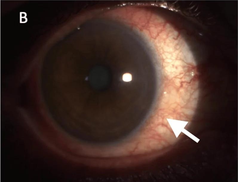
The arrow points to the inflamed sclera beside the cornea. After an attack settles, the sclera there often looks slightly grey-blue because it has become more translucent, not because it has thinned.
Nodular scleritis
Nodular scleritis forms one or more immobile nodules, usually near the limbus in the interpalpebral area, and the globe is tender. They are a deeper blue-red than an episcleral nodule, and 10% phenylephrine does not blanch the deep vessels over them.
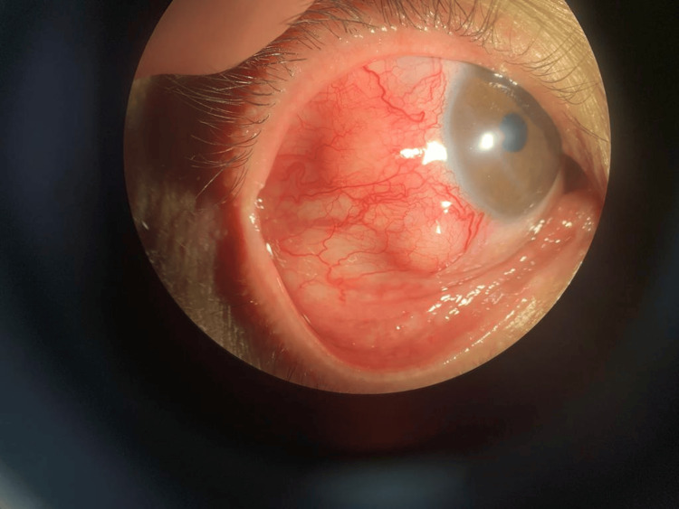
The nodule is raised from the sclera itself. More than 10% of patients with nodular scleritis go on to necrotizing disease, but early treatment prevents the necrosis.
Necrotizing scleritis
Necrotizing scleritis is the most destructive form. The pain is severe and persistent. Patches of sclera lose their blood supply and die, and as the sclera thins, the dark uvea shows through as a blue-black area. Patients tend to be older, and both eyes are often affected.
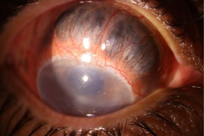
Above the cornea the sclera has thinned so much that the uvea shows through it.
Necrotizing scleritis can also be surgically induced. It starts at the site of a surgical wound, usually within 3 weeks but sometimes much later. It can follow trabeculectomy with mitomycin C, pterygium excision or a scleral buckle, and thinning can appear many years after a pterygium excision.
Scleromalacia perforans is a necrotizing scleritis without inflammation (about 5% of scleritis). It typically affects elderly women with long-standing rheumatoid arthritis. It is painless, and the eye is white, so the patient may only complain of mild irritation.
Posterior scleritis
Scleritis behind the equator is easy to miss because the eye may not be red. Pain is the most common symptom, vision is affected more often than in anterior scleritis, and anterior scleritis is present in only a minority. Many patients are young and otherwise healthy, but about a third of older patients have a systemic disease. It can look very like Vogt-Koyanagi-Harada disease, and the two are often hard to tell apart.
Look for:
- choroidal folds at the posterior pole
- exudative retinal detachment (about 25%) and macular edema
- choroidal detachment, disc swelling, mild proptosis, and pain on eye movement or double vision if the extraocular muscles are inflamed
- a shift toward hyperopia, as the thickened posterior sclera shortens the eye
Imaging confirms it. B-scan ultrasound shows a thickened sclera and fluid in the space under Tenon’s capsule. Where this fluid meets the optic nerve shadow, it forms the T-sign. The T-sign is very characteristic, but it is not always present.
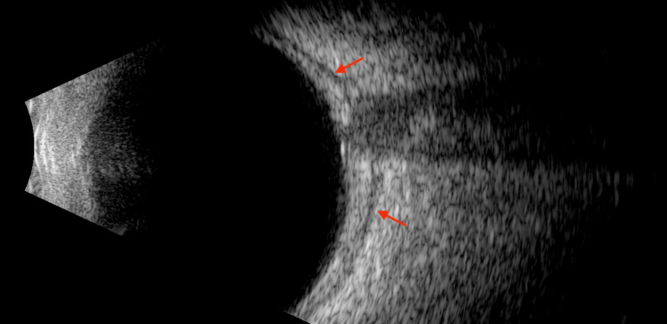
The red arrows mark the fluid along the back of the sclera. With the optic nerve shadow, it makes the shape of a T.
OCT may show subretinal fluid and a wavy, folded choroid. Vogt-Koyanagi-Harada disease gives the same exudative detachment and choroidal folds, and the fluorescein angiogram can leak in the same pattern. Here the T-sign on B-scan can settle the diagnosis. MRI and CT show the thickened sclera.
Inflammation can also spread forward from posterior scleritis and look like anterior scleritis. So when you see anterior scleritis, look at the back of the eye as well, with OCT and B-scan.
Uveitis and other complications
About one in four patients with scleritis has anterior uveitis, and in my experience anterior chamber cells are common. Severe uveitis may mean the scleritis is aggressive.
Glaucoma is the most common cause of permanent visual loss in anterior scleritis, so check and control the intraocular pressure. Scleritis next to the cornea can also cause keratitis, including peripheral ulcerative keratitis in necrotizing disease.
Treatment
Infection is unlikely, but always keep it in mind, and ideally do the systemic work-up before you start treatment.
For immune-mediated scleritis:
- Non-necrotizing scleritis: start with an oral NSAID. In my experience, celecoxib, a selective COX-2 inhibitor, works well. A COX-2 inhibitor may also be preferred in elderly patients or those with a history of peptic ulcer, but it can cause cardiovascular side effects. Topical steroids can ease the symptoms but do not change the course of the scleral inflammation. A periocular steroid injection can be used in non-necrotizing disease, but never in necrotizing scleritis.
- When NSAIDs are not enough, or the scleritis is necrotizing: systemic steroids, for example prednisolone 1 to 1.5 mg/kg daily. Severe cases can start with intravenous methylprednisolone 0.5 to 1 g daily for 3 days.
- Immunosuppressive drugs are added when steroids alone do not control the inflammation, to spare steroids, or to treat the underlying disease. Methotrexate is often used in rheumatoid arthritis. Patients with granulomatosis with polyangiitis, polyarteritis nodosa, relapsing polychondritis, or rheumatoid arthritis with necrotizing scleritis should start immunomodulatory treatment at once, together with the rheumatologist. Rituximab is particularly effective in necrotizing scleritis.
Infectious scleritis needs the specific antimicrobial treatment. Topical or systemic steroids may be added once the infection is being treated.
Summary
- Pain separates them: episcleritis is often painless, while scleritis usually gives a pain that wakes the patient at night.
- Phenylephrine blanches the superficial vessels but not the deep scleral plexus.
- Scleritis is often a systemic disease: work up rheumatoid arthritis and vasculitis, and think of infection.
- Necrotizing scleritis turns the sclera blue-black; when it is not infectious, it needs systemic steroids and immunosuppressive drugs.
- When you see anterior scleritis, look for posterior scleritis with OCT and B-scan.

Which retinal layer makes which wave, the three abnormal patterns, and the dark-adaptation trap to check before you report “negative” — on one page, by email. One email when I publish something new, about once a month. Unsubscribe anytime.