Peters Anomaly: Findings, Types I and II, and Management
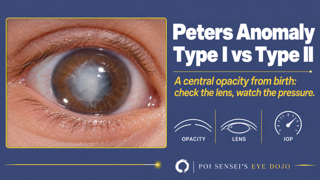
A newborn has a white opacity in the center of the cornea, and the peripheral cornea is relatively clear. Think of Peters anomaly. This article explains its cause, how to tell type I from type II, how to examine the eye, and what to watch for during follow-up.
- A central corneal opacity is present from birth.
- The peripheral cornea is relatively clear.
- Strands of iris run to the edge of the opacity.
- Both eyes are often affected.
What is Peters anomaly?
Peters anomaly is a congenital corneal opacity. During fetal development, neural crest cells fail to migrate normally, and the back of the central cornea does not form. The endothelium, Descemet membrane and posterior stroma are missing there. The corneal epithelium comes from the surface ectoderm, so it is normal. Only the tissues from the neural crest are missing.
About 80% of cases are bilateral. Most cases are sporadic, but families with recessive or dominant inheritance have been reported. Mutations in the PAX6 gene have been found in a few patients[1].
Type I
A central corneal opacity, usually with iridocorneal adhesions: strands of iris attach to the edge of the opacity. The lens is normal.
Type II
The features of type I plus lens abnormalities, such as cataract or a lens that has moved forward and sticks to the back of the cornea[2].
Findings
The opacity is in the center of the cornea, and its density varies from a faint haze to a dense white scar.
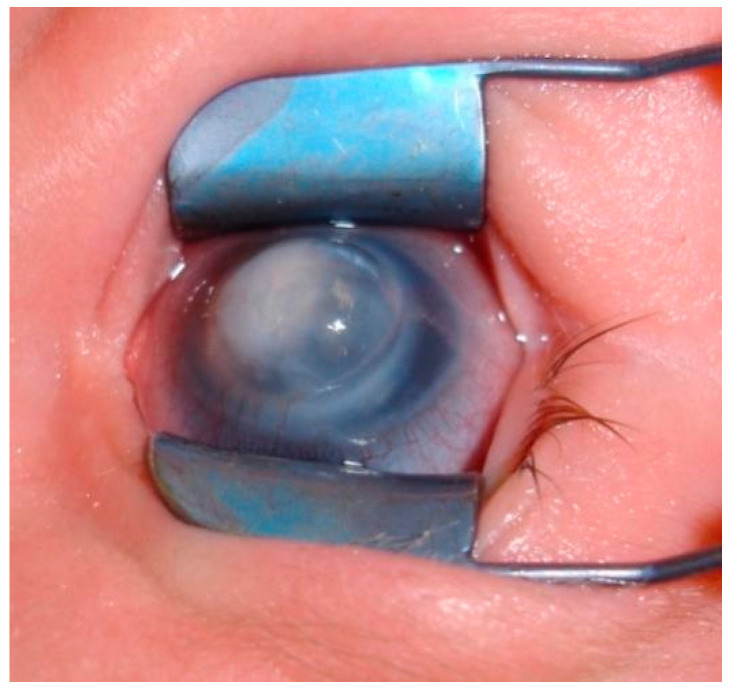
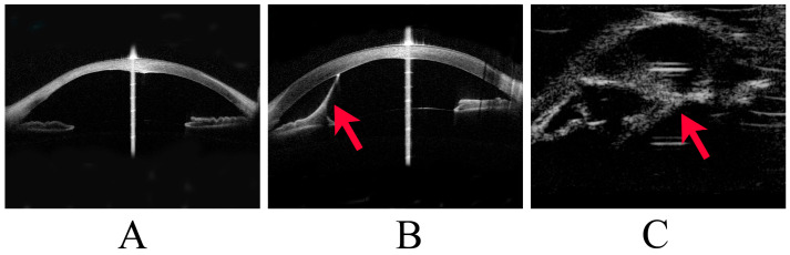
For the diagnosis, you need to see the back of the cornea, the iris and the lens. When the opacity is dense, the slit lamp cannot show them, but ultrasound biomicroscopy (UBM) and anterior segment OCT can. They show the posterior corneal defect, the iris strands, and whether the lens touches the cornea. In infants, the examination may need sedation or general anesthesia.
Complications
Eye
Peters anomaly is often the only anomaly in the eye, but because the neural crest forms many tissues of the eye, it can come with other anomalies: sclerocornea, coloboma, and persistent fetal vasculature (PFV, previously called persistent hyperplastic primary vitreous)[1].
Systemic
Some children also have anomalies outside the eye. When you suspect Peters anomaly from the eye findings, arrange a general pediatric examination. Reported associations include:
- short stature
- central nervous system anomalies and developmental delay
- cleft lip and palate
- heart defects
- pulmonary hypoplasia
- trisomy 13 or 15
Peters plus syndrome is an autosomal recessive form with short stature, developmental delay and cleft lip or palate.
Treatment
Corneal opacity
The opacity often becomes lighter as the child grows. The surrounding endothelium can slowly cover the central defect, and the corneal edema settles. Many children are therefore followed without surgery.
Even so, a central opacity in infancy can cause deprivation amblyopia, especially when only one eye is affected. Start amblyopia treatment early.
When a dense opacity blocks the visual axis in both eyes, penetrating keratoplasty is considered. If the opacity is small and the peripheral cornea is clear, an optical iridectomy (an opening in the iris behind the clear cornea) is a simpler option[1].
Glaucoma
Glaucoma develops in about half of patients, in both type I and type II. It usually starts in infancy, but it can appear later in childhood[2].
Follow and control the intraocular pressure (IOP) from the first visit. If eye drops do not control the IOP, surgery such as trabeculotomy is done. If glaucoma is present, treat it before any corneal surgery.
Related conditions
Peters anomaly is one of the anterior segment dysgenesis disorders.
Axenfeld-Rieger syndrome
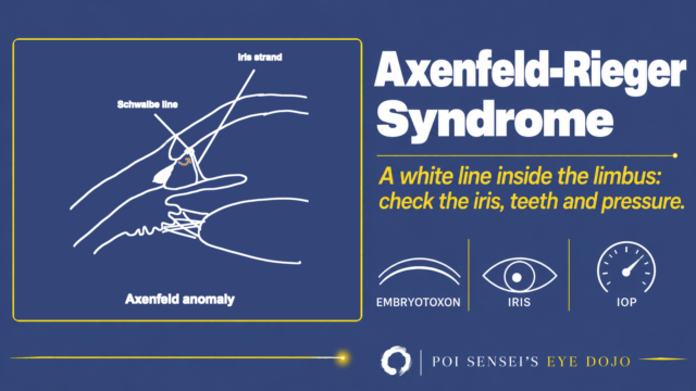
The Schwalbe line is thick and displaced forward (posterior embryotoxon), and strands of iris attach to it. Glaucoma develops in about half of patients. Dental and facial anomalies may also be present[2].
Sclerocornea
The cornea turns white like the sclera, either at the periphery or over the whole cornea. It can occur together with Peters anomaly[1].
Summary
- Peters anomaly is a congenital central corneal opacity from abnormal neural crest migration. About 80% are bilateral.
- Type I: central opacity with iris strands. Type II: type I plus lens abnormalities.
- UBM and anterior segment OCT show the back of the cornea, the iris and the lens.
- The opacity often becomes lighter with growth, but amblyopia treatment should start early.
- Glaucoma occurs in about half of patients. Follow and control the IOP.
Figure credits
Clinical photograph: Figure 2 from Wowra B, Dobrowolski D, Parekh M, et al. J Clin Med 2024;13(2):532 (https://doi.org/10.3390/jcm13020532), licensed under CC BY 4.0. Used without changes.
Anterior segment OCT and UBM: Figure 1 from Yokota C, Hirooka K, Okada N, et al. J Clin Med 2023;12(19):6238 (https://doi.org/10.3390/jcm12196238), licensed under CC BY 4.0. Used without changes.
References
[1] Yanoff M, Duker JS, eds. Ophthalmology. 5th ed. Elsevier; 2019. Chapter 4.3, Congenital corneal anomalies.
[2] Salmon JF. Kanski’s Clinical Ophthalmology: A Systematic Approach. 10th ed. Elsevier; 2025. Chapter 11, Glaucoma: iridocorneal dysgenesis.
Which retinal layer makes which wave, the three abnormal patterns, and the dark-adaptation trap to check before you report “negative” — on one page, by email. One email when I publish something new, about once a month. Unsubscribe anytime.