Infantile Nystagmus: Five Early-Onset Types and How to Tell Them Apart
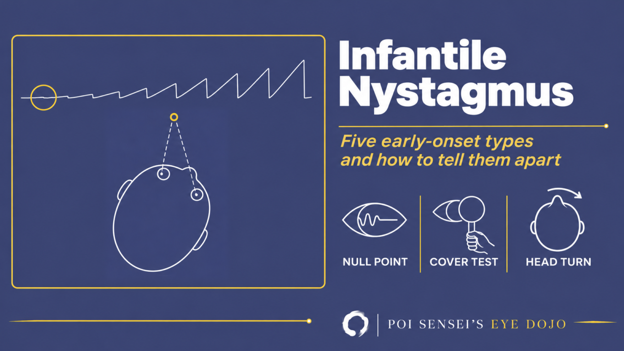
Nystagmus that starts in the first months of life is called early-onset or infantile nystagmus. It is not one disease. There are five main types: infantile nystagmus syndrome, periodic alternating nystagmus, latent nystagmus, nystagmus blockage syndrome and spasmus nutans. Some overlap: nystagmus blockage syndrome, for example, starts from infantile nystagmus syndrome.
This article covers how to tell them apart, what to check next, and when to order an MRI.
Infantile nystagmus syndrome
The most common type is infantile nystagmus syndrome (INS), once called congenital nystagmus. Despite the old name, it is rarely present at birth. It usually shows up at around 3 months of age.
Its features are:
- The two eyes move together, by about the same amount, and the nystagmus is horizontal. It stays horizontal even on up- and downgaze.
- It gets worse when the child tries hard to fixate and quieter with convergence. It stops during sleep.
- Oscillopsia is rare. The world does not seem to move, even for most adults with INS.
- Acuity is reduced, typically to around 20/40 to 20/120, and lower still with an eye disease such as albinism.
- Refractive errors, especially astigmatism, are common.
Sensory or motor
INS falls into two groups.
In sensory INS, poor central vision from early life stops the eyes from holding steady. Causes include albinism, aniridia, congenital cataract, optic nerve hypoplasia, Leber congenital amaurosis and achromatopsia. The movement is often pendular: the eyes swing back and forth at the same speed, with no fast phase. This is the more common group.
In motor INS, the visual pathway is normal. It often runs in families, most often X-linked. Vision is usually better than in the sensory group.
A child with typical INS, normal development, a normal fundus and a normal ERG and VEP usually does not need brain imaging first. Neither does a child with a clear eye cause, such as albinism or a retinal dystrophy. Nystagmus in one eye only, or very asymmetric nystagmus, is different: image the brain[1].
Null point and head turn
The nystagmus changes over the first years. Large, slow swings in early infancy become pendular, and later a jerk nystagmus: a slow drift followed by a fast flick back. A jerk nystagmus is named by the direction of its fast phase.
Most children then settle on a null point: a direction of gaze where the nystagmus is weakest and vision is best. The further the eyes look away from it, the stronger the nystagmus becomes. To keep the eyes at the null point while looking straight ahead, the child turns the face.
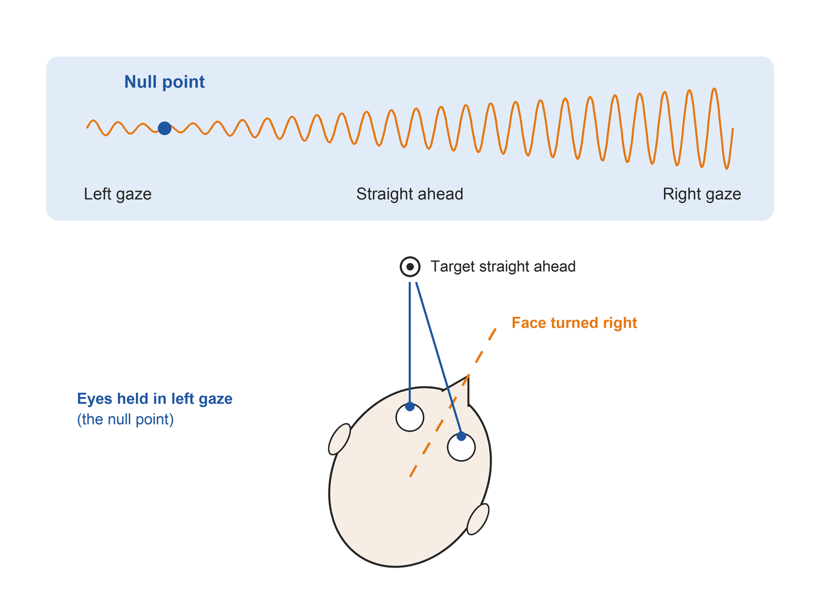
In the top row, the nystagmus is weakest in left gaze and strongest in right gaze, so the null point lies in left gaze. In the bottom row, the child turns the face to the right. The eyes then sit in left gaze, at the null point, while looking at a target straight ahead.
A classic sign is a reversed optokinetic response: with an optokinetic nystagmus (OKN) drum, the eyes beat the opposite way from normal. In clinic, though, it is often hard to see any clear horizontal OKN on top of the nystagmus[1].
Treatment
First, correct the refractive error, astigmatism included.
Prisms can help. Base-out prisms make the eyes converge, which dampens the nystagmus. Prisms with their apex toward the null point move the eyes toward it without a face turn.
If the face turn is large, surgery can move the null point to straight ahead (the Kestenbaum–Anderson procedure). Take the child with the null point in left gaze. Recessing the right medial rectus and the left lateral rectus shifts both eyes to the right. To look straight ahead, the brain now has to send the same command as for left gaze, so the eyes sit at the null point with the face straight. The null point can drift again months after surgery.
Other early-onset types
Periodic alternating nystagmus
Periodic alternating nystagmus (PAN) is a horizontal jerk nystagmus that changes direction every minute or two. It can be congenital, often with albinism, or acquired from a lesion of the cerebellum. Unlike INS, it often causes oscillopsia.
It is thought to come from a loss of GABA-mediated control in the cerebellum, and baclofen, which acts on GABA receptors, reduces it.
Watch the eyes for at least 5 minutes, ideally 10[1]. If the face turn seems to change from visit to visit, suspect PAN. Large recessions of all four horizontal rectus muscles can reduce the nystagmus and the head turn.
Latent nystagmus
With both eyes open, there is no nystagmus. Cover one eye, and a jerk nystagmus appears with the fast phase toward the uncovered eye. Cover the other eye, and the direction reverses. When one eye sees poorly, the nystagmus can show even with both eyes open (manifest latent nystagmus).
The current name for both is fusion maldevelopment nystagmus syndrome (FMNS). It goes with infantile esotropia, where it is found in about half of patients, and with dissociated vertical deviation.
Because covering one eye brings out the nystagmus, binocular acuity is better than monocular acuity. To measure each eye, blur the other eye with a plus lens instead of covering it.
Nystagmus blockage syndrome
A child with INS can damp the nystagmus by converging. If the child keeps converging, the eyes stay turned in, and an esotropia appears. The child fixates with the adducted eye and turns the face so that eye can see the target.
It has to be told apart from infantile esotropia and accommodative esotropia. Two clues point to nystagmus blockage: the nystagmus appears when the fixing eye moves out into abduction, and the angle of the esotropia varies. After strabismus surgery, the esotropia tends to come back, because the child keeps converging to block the nystagmus.
Spasmus nutans
Spasmus nutans starts between about 3 and 18 months of age. Its triad is nystagmus, head nodding and an abnormal head posture. The nystagmus is small, fast and often different between the two eyes, sometimes in one eye only. The head nodding is thought to help cancel it.
Most cases resolve on their own by about age 3. Strabismus and amblyopia can occur alongside it, so treat amblyopia when needed.
The same picture can come from a glioma of the anterior visual pathway, and the eye movements alone cannot reliably tell them apart. Some clinicians image every child, as with any nystagmus in one eye. If there is vision loss, optic atrophy, delayed development or any other neurological sign, always order an MRI.
Summary
- Early-onset nystagmus comes in five main types; INS is the most common.
- INS is horizontal, gets worse with fixation and quieter with convergence, and rarely causes oscillopsia.
- Look for an eye cause with OCT and ERG; image the brain for one-eyed nystagmus or neurological signs.
- Latent nystagmus beats toward the uncovered eye, PAN changes direction every minute or two, and spasmus nutans adds head nodding.
References
[1] Osborne D, Theodorou M, Lee H, et al. Supranuclear eye movements and nystagmus in children: A review of the literature and guide to clinical examination, interpretation of findings and age-appropriate norms. Eye (Lond). 2019;33(2):261–273. PMID 30353137.
Which retinal layer makes which wave, the three abnormal patterns, and the dark-adaptation trap to check before you report “negative” — on one page, by email. One email when I publish something new, about once a month. Unsubscribe anytime.