Central Retinal Artery Occlusion (CRAO): The First Hour
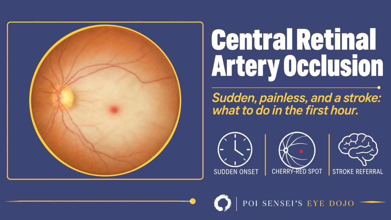
Central retinal artery occlusion (CRAO) is a stroke in the eye: the artery that feeds the inner retina is suddenly blocked. A branch retinal artery occlusion (BRAO) differs mainly in how much retina is lost, so this article follows CRAO, how to recognize it and what to do in the first hour, and covers BRAO briefly at the end.
Sudden, not just acute
Ask the patient: "When did the vision go, and what were you doing?" A patient with CRAO can usually answer exactly, down to the minute: watching television, standing up from a chair. That is sudden onset. A patient with optic neuritis cannot. Their vision got worse over hours or days, so they can only say "since this morning" or "over the weekend". The answer alone is not a diagnosis. Still, it narrows the list. Sudden loss of vision usually means a blocked or a burst vessel: CRAO is a blocked one, a vitreous hemorrhage a burst one.
The loss of vision is painless, except in giant cell arteritis, and it is severe. Vision is often counting fingers or worse. If there is no light perception, think of giant cell arteritis or an occlusion of the whole ophthalmic artery. The relative afferent pupillary defect (RAPD) is marked and sometimes total. Some eyes have a cilioretinal artery, a branch of the ciliary circulation that supplies part of the macula. If it reaches the fovea, central vision can survive.
Why the retina turns white and the fovea stays red
The retina has two blood supplies. The inner retina is fed by the retinal vessels, from the central retinal artery. The outer retina, including the photoreceptors, is fed by the choroid. When the central retinal artery is blocked, only the inner retina loses its blood. It swells and becomes opaque, so the posterior pole looks white.
The fovea is different. At the foveal center there is no inner retina, only the outer layers. Nothing there swells, and the orange-red of the healthy choroid shows through. That is the cherry-red spot.
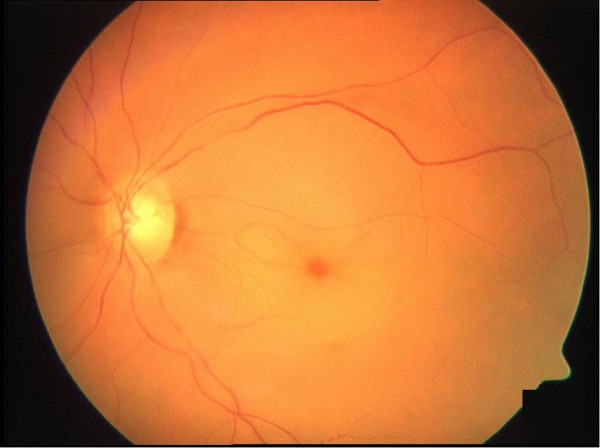
In this left eye the retina around the macula is pale, the arteries are narrow, and the fovea stands out as a small red spot.
Other signs help. The blood column in the arteries and veins can break into segments (boxcar segmentation). An embolus is visible in about 20% of eyes. Unlike retinal vein occlusion, there are usually no hemorrhages and the veins are not dilated.
Timing matters when you look. The whitening can take several hours to develop, so an eye seen very early can look almost normal. Over days to weeks the whitening and the cherry-red spot fade. The arteries stay narrow, and the disc becomes pale.
OCT, FA and ERG
All three follow the same rule: the inner retina is damaged and the outer retina is spared. In the acute phase, do them only if they do not delay the referral described below.
OCT
In the acute phase the inner retina is thick and hyperreflective, and the layers below it look darker in its shadow. Because the retina around the fovea thickens and the foveal center does not, the foveal pit can look deeper than normal. The earliest change, before the full picture, may be a hyperreflective band in the inner nuclear layer, called paracentral acute middle maculopathy (PAMM).
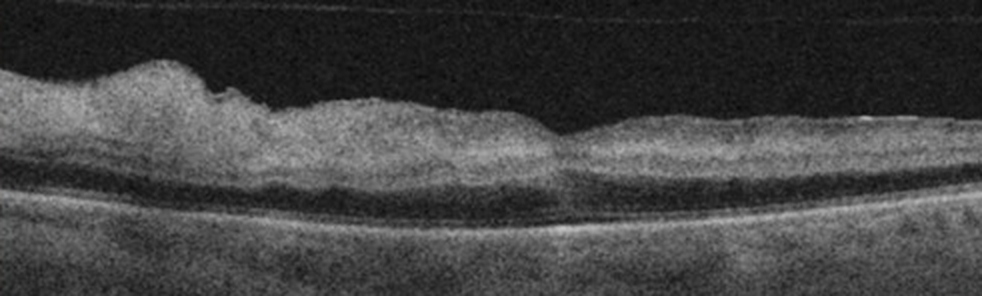
Here the inner retina is swollen and bright, while the outer retina and the retinal pigment epithelium below are intact.
Over the following weeks the inner retina thins. The outer layers stay, and the foveal pit becomes flat. See how to read a macular OCT for the layers.
Fluorescein angiography
Fluorescein angiography shows delayed filling. The time from the arm injection to the retinal arteries is long, and so is the time from the arteries to the veins. Even when the arteries look filled, these delays still show the occlusion. A patent cilioretinal artery fills early.
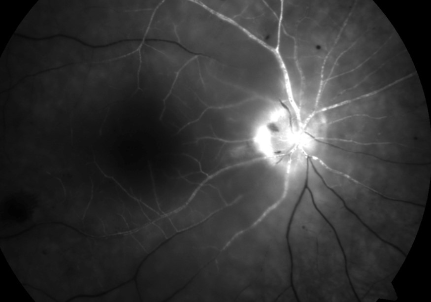
In this right eye the arteries have filled with dye, but the veins are still dark: the dye is moving slowly through the retina.
ERG
The bipolar and amacrine cells sit in the inner retina, so the b-wave falls and the oscillatory potentials disappear, while the photoreceptors still give an a-wave. The result is a negative ERG. You rarely need it for CRAO, but it helps when the fundus signs are subtle and you are unsure whether the problem is in the retina or the optic nerve. The ERG article explains the waves.
The first hour
Once you suspect CRAO, the next steps are not in the eye clinic.
Treat it as a stroke
The central retinal artery is a branch of the ophthalmic artery, the first branch of the internal carotid artery, so an embolus from a carotid plaque has an easy route to the eye. CRAO shares its causes with brain stroke, and the guidelines manage it the same way. The risk of stroke is highest around the event, from about 2 weeks before to 1 month after. About a fifth of patients with acute retinal artery occlusion already have a silent infarct on diffusion-weighted MRI[1].
So refer the patient immediately to an emergency department with a stroke service, and say clearly that this is an eye stroke. Tests such as FA or OCT should not delay the transfer.
Check for giant cell arteritis
Giant cell arteritis causes only about 1 to 2% of CRAO, but it can blind the other eye. In anyone over 50, ask about headache, jaw claudication, scalp tenderness, weight loss and shoulder or hip girdle pain, and order ESR, CRP and a platelet count. Giant cell arteritis typically shows disc swelling and no visible embolus. If you strongly suspect giant cell arteritis, start systemic steroids now and arrange the temporal artery biopsy afterwards. Normal ESR and CRP do not completely rule it out.
Thrombolysis
Intravenous thrombolysis within 4.5 hours, as in a brain stroke, has been tried because the retina can recover only if blood flow returns within a few hours. Two randomized trials have now compared it with aspirin within 4.5 hours. Neither alteplase, in a small trial[2], nor tenecteplase[3] gave a significant gain in vision, and tenecteplase caused more serious adverse events, including one fatal intracranial hemorrhage. So thrombolysis has not been shown to restore vision. Whether to offer it is a decision for the stroke team.
Eye treatments
Ocular massage, anterior chamber paracentesis and lowering the intraocular pressure have all been tried. None has been shown to do better than the natural course, and a meta-analysis suggested that eyes given these treatments may even do worse. Most eyes keep poor vision: about two-thirds end worse than 20/400 (6/120), and only about a fifth reach 20/40 (6/12) or better.
Finding the cause
Most patients are older and have hypertension, diabetes or other vascular risk factors. The usual sources are an embolus from a carotid plaque or from the heart, especially with atrial fibrillation. Less common causes are carotid dissection (ask about neck pain or recent trauma), vasculitis, antiphospholipid syndrome and blood disorders.
Much of the work-up is done by the stroke team, and it is a stroke work-up: blood pressure, pulse and ECG for arrhythmia, carotid duplex ultrasound, and blood tests including glucose, lipids and a full blood count. Echocardiography is usually added in younger patients or when the heart is a likely source. In younger patients or without vascular risk factors, look further for thrombophilia, antiphospholipid antibodies and vasculitis. Even after a full work-up, the cause often stays unknown.
Afterwards: watch for new vessels
Iris new vessels develop in up to about 1 in 5 eyes, typically 4 to 5 weeks after onset. That is earlier than in central retinal vein occlusion, where they usually appear at about 3 months. New vessels on the retina or the disc are less common, about 2%.
See the patient at 3 to 4 weeks, and then at least twice more at monthly intervals. At each visit look at the pupil margin and do gonioscopy before dilating, because new vessels in the angle can appear first. Do not apply panretinal photocoagulation (PRP) in advance. Apply it when new vessels appear, with an anti-VEGF injection as an option.
Branch retinal artery occlusion
In BRAO the signs are the same but limited to the area of the blocked branch. The patient notices a sudden sectoral or altitudinal field defect. If central vision is spared, the patient may not notice it at all. Look for an embolus at an arterial bifurcation. The field defect rarely recovers. An acute, symptomatic BRAO needs the same urgent stroke referral as CRAO. An asymptomatic BRAO, or an embolus found by chance in a patient with no symptoms, can go to the primary care doctor for a vascular work-up.
Summary
- CRAO starts suddenly, so ask when the vision went and what the patient was doing.
- The inner retina turns white and the fovea stays red, but very early the fundus can look normal.
- Refer the patient immediately as a stroke. Over 50, check ESR and CRP, and start steroids if you strongly suspect giant cell arteritis.
- Thrombolysis and eye treatments have not been shown to restore vision.
- Look for iris and angle new vessels in the first months, and apply PRP when they appear.
References
[1] Kovach JL, Bailey ST, Kim SJ, et al. Retinal and Ophthalmic Artery Occlusions Preferred Practice Pattern. Ophthalmology. 2025;132(4):P270-P302. PMID 39918522.
[2] Préterre C, Gaultier A, Obadia M, et al. Intravenous alteplase versus oral aspirin for acute central retinal artery occlusion within 4·5 h of severe vision loss (THEIA): a multicentre, double-dummy, patient-blinded and assessor-blinded, randomised, controlled, phase 3 trial. Lancet Neurol. 2025;24(11):909-919. PMID 41109232.
[3] Ryan SJ, Jørstad ØK, Skjelland M, et al. A Randomized Trial of Tenecteplase in Acute Central Retinal Artery Occlusion. N Engl J Med. 2026;394(5):442-450. PMID 41604638.

Which retinal layer makes which wave, the three abnormal patterns, and the dark-adaptation trap to check before you report “negative” — on one page, by email. One email when I publish something new, about once a month. Unsubscribe anytime.