Branch vs Central Retinal Vein Occlusion: Where the Vein Is Blocked
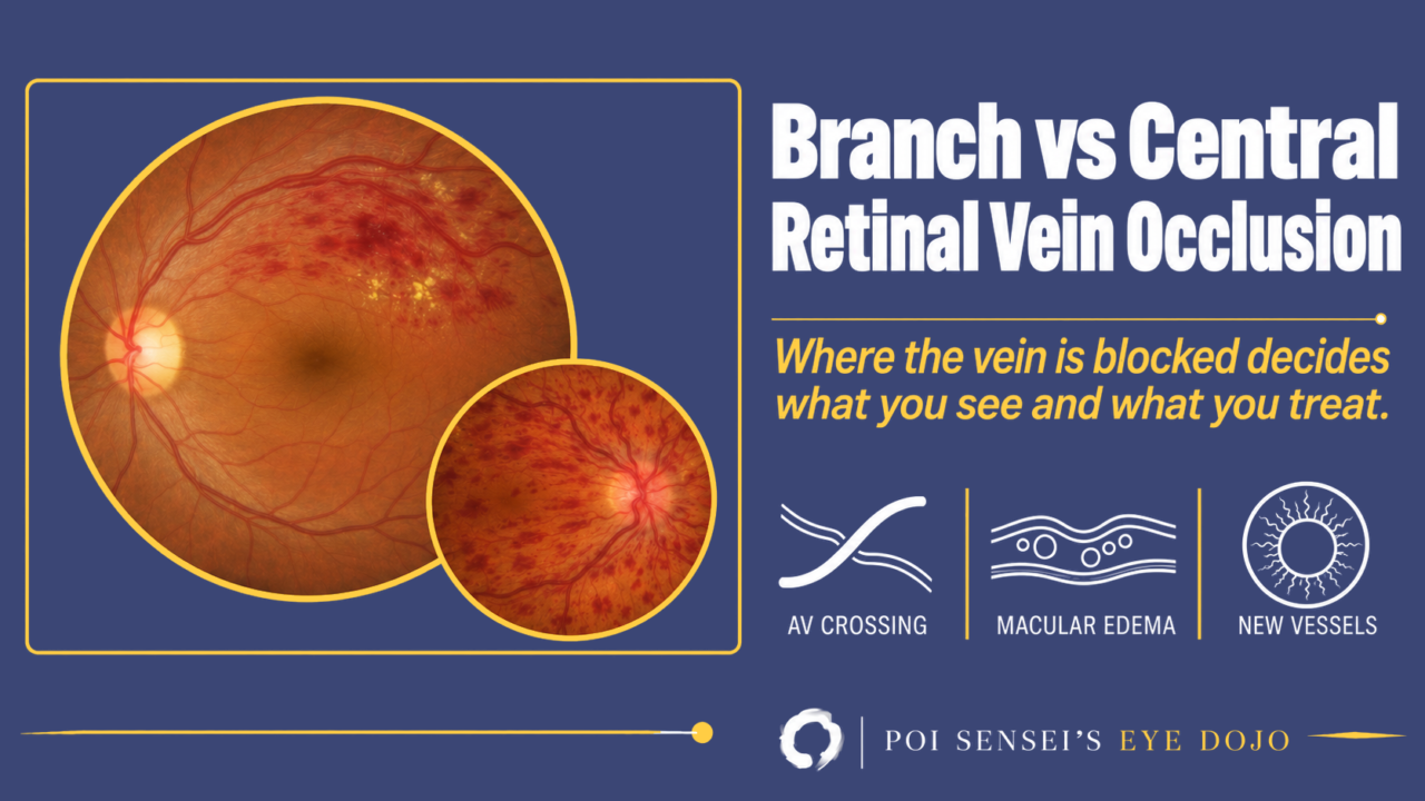
Retinal vein occlusion (RVO) is a blockage of a retinal vein, and it divides into two main types: branch retinal vein occlusion (BRVO) and central retinal vein occlusion (CRVO). RVO is the second most common retinal vascular disease after diabetic retinopathy. It is tempting to think of CRVO as a severe BRVO, but the two differ in where and why the vein is blocked, and that changes what you see, which test matters and what you treat.
Where the vein is blocked
In BRVO the block is at an arteriovenous crossing. There, a retinal arteriole and venule share a sheath, and an arteriole thickened by arteriosclerosis presses on the venule. The flow becomes turbulent and a thrombus forms. In CRVO the central retinal vein is blocked inside the optic nerve, near the lamina cribrosa, where it also shares a sheath with the central retinal artery.
The background is mostly the same. Age is the most important risk factor. Hypertension is common and matters most in BRVO. Hyperlipidemia is common too, and glaucoma raises the risk of CRVO. So in every patient, check the blood pressure, blood glucose and lipids, with a full blood count and ESR. In a patient under 50, in a bilateral occlusion, or after a previous thrombosis, look further for blood and inflammatory disorders, such as thrombophilia and autoimmune disease. A woman taking the contraceptive pill should probably stop it. Antiplatelet drugs and anticoagulants are not usually started for the occlusion itself, only when there is another systemic reason.
The hemorrhages look different depending on their depth. A hemorrhage in the superficial nerve fiber layer spreads along the nerve fibers and looks flame-shaped. A hemorrhage in the deeper, middle layers cannot spread, so it looks round, like a dot or blot. The venous ends of the capillaries lie in the middle layers, so when venous pressure rises, those small vessels break first. That is why a mild occlusion shows mostly blot hemorrhages, and flame hemorrhages appear as it gets worse.
Branch retinal vein occlusion
The signs are only in the area drained by the blocked vein, distal to the crossing. The vein is dilated and tortuous, with flame and blot hemorrhages, sometimes with cotton-wool spots and edema. The superotemporal branch is the most common. If the macula is involved, the patient notices sudden blurred or distorted vision. A peripheral BRVO can go unnoticed.
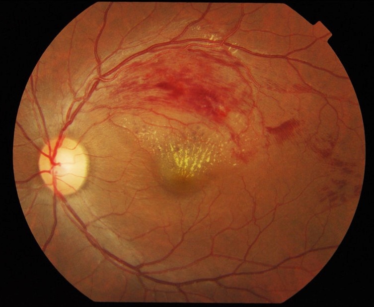
In this left eye the hemorrhages and hard exudates sit in the upper temporal sector only, and the lower half of the fundus is normal.
Chronic macular edema is the most common cause of poor vision in BRVO, so OCT is the key test. If there is center-involving macular edema, consider treatment. Even when visual acuity is good, take an OCT if the patient notices distortion or vague blurring.
- Intravitreal anti-VEGF injection is the usual first choice. In Japan a common approach is one injection, then another each time the edema returns (1+PRN). In trials, monthly injections came first, and the gains held with as-needed treatment later.
- A dexamethasone implant is an alternative. It can be repeated after 4 to 6 months, but it raises the risk of high intraocular pressure and cataract.
- Macular laser is now an add-on. Consider it when edema keeps vision at 20/40 (6/12) or worse after 3 to 6 months and FA shows good perfusion at the macula.
FA matters less in BRVO than in CRVO. It shows the non-perfused area, but non-perfusion alone is not a reason for laser. Retinal new vessels develop in about 8% of eyes within 3 years, usually in the first 6 to 12 months, and the risk is higher when non-perfusion exceeds about five disc areas. Even then, watch, and apply sector laser to the non-perfused area when new vessels appear. Iris new vessels are rare in BRVO, but if they appear, sector laser is urgent. Review at 3 months, then every 3 to 6 months for up to 2 years, mainly to catch new vessels. Widefield OCT angiography can also show the non-perfused area without dye.
Over weeks to months, collateral vessels may form, often crossing the horizontal raphe. They are a good sign, so do not treat them with laser.
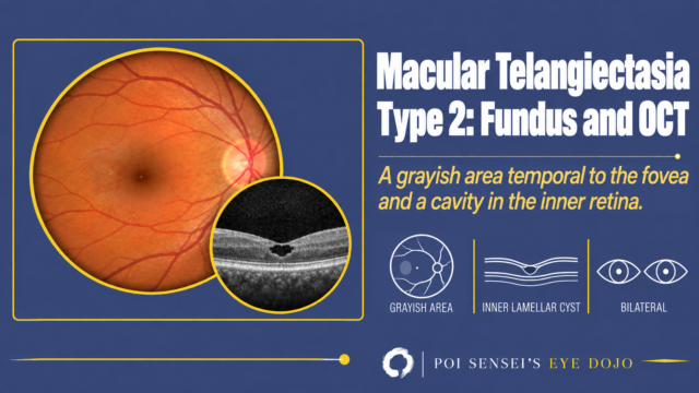
A small BRVO near the macula can look like other causes of macular leakage. On FA, check whether the leakage starts beyond an arteriovenous crossing. If it does, suspect a BRVO. If the leakage has nothing to do with a crossing, consider macular telangiectasia or Coats disease.
Central retinal vein occlusion
The signs are in all four quadrants: every branch of the central retinal vein is dilated and tortuous, with flame and blot hemorrhages, cotton-wool spots and often disc and macular edema. Vitreous hemorrhage can follow.
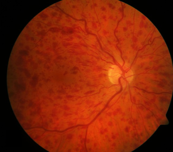
In this right eye the hemorrhages reach every quadrant, around the disc and out to the periphery, and the veins are dilated and tortuous.
In CRVO, first decide whether it is ischemic or non-ischemic. The classic definition comes from the Central Vein Occlusion Study (CVOS): more than 10 disc areas of capillary non-perfusion on FA means ischemic CRVO. Widefield imaging sees much more of the periphery, so this cut-off fits it less well; what still holds is that more than 10 disc areas of non-perfusion at the posterior pole means a high risk of new vessels. Before angiography, the clinical picture already helps. Vision worse than 20/200 (6/60) with a relative afferent pupillary defect usually means significant ischemia, as do many dark, deep blot hemorrhages and cotton-wool spots. These are not the few small blots of a mild occlusion: in ischemic CRVO the deep hemorrhages are extensive.
Non-ischemic CRVO is the more common form. About a third becomes ischemic, often within months, so it needs follow-up even when it looks mild. See the patient again after 3 months, and ask them to come back sooner if vision drops or the eye becomes painful or red, because that can mean ischemia or neovascular glaucoma.
In ischemic CRVO, iris new vessels develop in about half of eyes, usually 2 to 4 months after onset, so it was once called "100-day glaucoma". At each visit, look at the pupil margin and do gonioscopy before you dilate the pupil, because new vessels in the angle can appear without any at the pupil margin. See ischemic CRVO monthly for the first 6 months, then keep watching for 2 to 3 years. Once disc collaterals (optociliary shunts) form, the risk of new vessels falls.
Treatment follows two lines: one for macular edema and one for new vessels.
If there is center-involving macular edema, consider treatment, and start early: delaying treatment by up to 6 months gives smaller gains. Use intravitreal anti-VEGF or a dexamethasone implant.
- In Japan, three monthly injections and then as needed (3+PRN), or 1+PRN, are common. The UK guideline gives monthly injections until vision is at its best and stable, then treats as needed with a review every 4 to 8 weeks.
- Even when vision is 20/320 (6/96) or worse, anti-VEGF is still worth trying if the edema is marked. If the edema resolves after 3 injections but vision does not improve, stop.
- Unlike in BRVO, macular laser does not improve vision in CRVO.
For new vessels: if iris or angle new vessels appear, apply panretinal photocoagulation (PRP) without delay. The UK guideline also gives an anti-VEGF injection at the first sign, with the PRP on the same day, before the injection, or within 1 to 2 weeks. Whether to apply PRP before new vessels appear is debated. Kanski finds no benefit[1], but the UK guideline says it can be considered when non-perfusion at the posterior pole exceeds 10 disc areas[2].
Anti-VEGF given for the edema does not remove the risk of new vessels. After the injections stop, check an ischemic CRVO for new vessels every 1 to 2 months for the first year. With a dexamethasone implant, look for iris new vessels at every visit, so you catch them as early as possible.
The ERG reflects how much of the inner retina is ischemic. The inner retina is supplied by the retinal circulation and the outer retina by the choroid. In milder cases, only the amacrine cells drop out, and the oscillatory potentials (OPs) disappear. In ischemic CRVO the bipolar cells are damaged too, so the b-wave falls while the a-wave from the photoreceptors is preserved. The result can be a negative ERG, with the b-wave smaller than the a-wave. ERG is seldom needed; when it is recorded, a low b-wave supports ischemic CRVO.
Three other forms of CRVO
- Hemiretinal vein occlusion: some people have two trunks of the central retinal vein. If one is blocked, only the upper or lower half of the retina is affected, with an altitudinal field defect. If ischemia is extensive, treat it like ischemic CRVO; otherwise, macular edema is managed as in BRVO.
- Impending CRVO: a mild form with slight venous dilatation and a few blot hemorrhages. There is no non-perfusion, and macular edema, if any, is mild. Vision may be only slightly blurred, often worse on waking. There is no proven treatment. If there is macular edema, treat it as in non-ischemic CRVO; if not, observe carefully. The prognosis is usually good, but some eyes progress to ischemic CRVO.
- CRVO in younger patients: CRVO can occur under 50 without hypertension or other vascular risk factors. Disc swelling often dominates, with hemorrhages mostly around the disc, and inflammation of the disc or its vessels may be involved; this is sometimes called papillophlebitis. Vision is usually only mildly to moderately reduced, without an RAPD, and the prognosis is generally good. If both eyes are affected, rule out raised intracranial pressure. Investigate it as you would any CRVO in a young patient. If the disc or its vessels are inflamed, consider steroids once systemic causes are excluded: oral prednisolone at about 0.6 to 0.7 mg/kg/day, tapered over several months. If this helps too little or the occlusion worsens during the taper, consider a sub-Tenon triamcinolone injection or intravitreal anti-VEGF.
OCT after treatment
OCT shows more than whether the edema is gone. Check whether the ellipsoid zone is continuous. Also look for the foveal bulge, the small upward bump of the ellipsoid zone under the fovea. When vision stays poor even though the edema has resolved and the ellipsoid zone looks intact, the foveal bulge is often missing.
Summary
- BRVO is blocked at an arteriovenous crossing and affects one sector; CRVO is blocked inside the optic nerve and affects all four quadrants.
- In both, treat macular edema; in BRVO, laser for non-perfusion waits until new vessels appear.
- In CRVO, decide ischemic or non-ischemic. Watch the iris and the angle for new vessels, and treat them with PRP without delay.
- In every patient, check blood pressure, blood glucose and lipids, and look further in younger patients.
References
[1] Salmon JF. Kanski’s Clinical Ophthalmology: A Systematic Approach. 10th ed. Elsevier; 2025. Chapter 13, Retinal vascular disease: central retinal vein occlusion.
[2] The Royal College of Ophthalmologists. Clinical Guidelines: Retinal Vein Occlusion (RVO). London: RCOphth; 2022.

Which retinal layer makes which wave, the three abnormal patterns, and the dark-adaptation trap to check before you report “negative” — on one page, by email. One email when I publish something new, about once a month. Unsubscribe anytime.